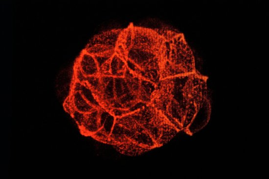
© Hugues LELOUARD / CIML / INSERM / CNRS Images
Reference
20240015_0003
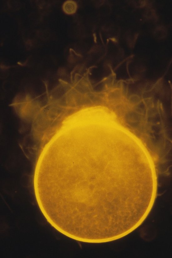
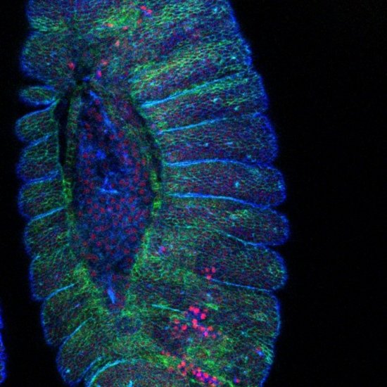
Coupe d'intestin grêle d'une souris transgénique contenant une plaque de Peyer
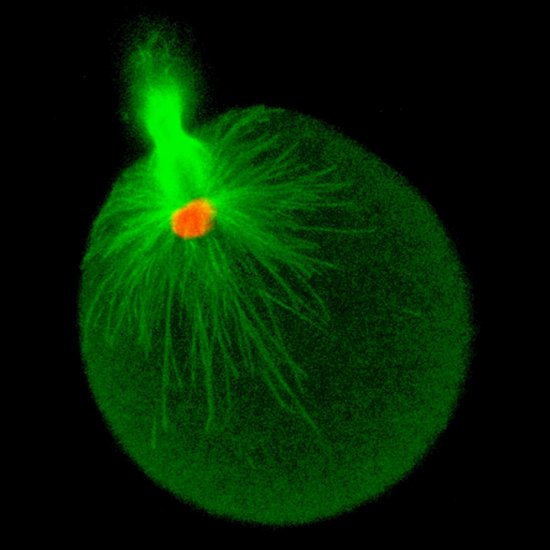
Small intestine section from a LyzM-eGFP mouse containing one Peyer’s patch and stained for proliferative cells (Ki-67, yellow), Paneth cells (UEA-I, blue), epithelial cells (EpCAM, magenta), naive B cells (IgD, red), T cells (CD3, orange), helper T cells/macrophages (CD4, cyan), phagocytes (CD11c, turquoise), monocyte-derived phagocytes (GFP, green). This image is the winner of the 2023 edition of the France-Bioimaging image competition (UMS3714-CEMIBIO, CNRS).
Add to my selection
Terms of use
The use of media visible on the CNRS Images Platform can be granted on request. Any reproduction or representation is forbidden without prior authorization from CNRS Images (except for resources under Creative Commons license).
No modification of an image may be made without the prior consent of CNRS Images.
No use of an image for advertising purposes or distribution to a third party may be made without the prior agreement of CNRS Images.
For more information, please consult our general conditions