© Mégane RAYER / LBCMCP / CNRS Images
Reference
20170079_0007
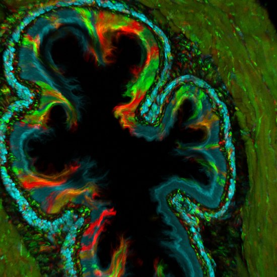
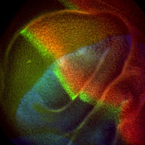
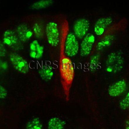
Cellule épithéliale en phase d’apoptose observée en microscopie confocale
Epithelial cell in the developing leg of a fruit fly, during apoptosis (programmed cell death), observed using a confocal microscope. The cell nuclei are visible in green. The aim of research scientists is to understand how programmed cell death can influence how tissues change shape.
The use of media visible on the CNRS Images Platform can be granted on request. Any reproduction or representation is forbidden without prior authorization from CNRS Images (except for resources under Creative Commons license).
No modification of an image may be made without the prior consent of CNRS Images.
No use of an image for advertising purposes or distribution to a third party may be made without the prior agreement of CNRS Images.
For more information, please consult our general conditions
