Production year
2011

© Jonathan NOWAK / Clément GHIGO / Marc BAJENOFF / INSERM / CIML / CNRS Images
20170103_0018
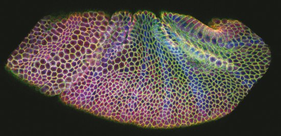
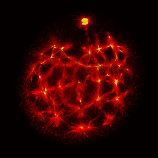
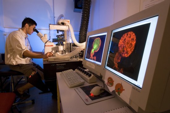
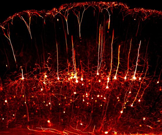
Mouse oesophagus cells, imaged using confocal microscopy. This image of an oesophagus was acquired in a mouse that had been genetically modified to randomly express various colours in each of its cells, thereby differentiating one cell from another. The image was obtained by combining several lasers, including a white-light laser that generates a wide range of excitation wavelengths, enabling multiple colour fluorescent proteins to be imaged. A high-power oil-immersion objective lens was used. With confocal microscopy, laser beams scan the specimen at high speed, point by point. This technique can be used to produce thin optical cross-sections (at different altitudes in a thick specimen) and very precisely indicate the locations of numerous entities and cell types in a tissue slice. These optical cross-sections are then stacked to generate 3D images of the specimens. This enables scientists to compare the locations and numbers of entities in various circumstances, such as in patients suffering from diseases, for example, in order to shed light on the workings of the immune system. This technique is now widely used in laboratories, and is constantly being improved in terms of its resolution and sensitivity, as well as the possible colours and contrasts.
The use of media visible on the CNRS Images Platform can be granted on request. Any reproduction or representation is forbidden without prior authorization from CNRS Images (except for resources under Creative Commons license).
No modification of an image may be made without the prior consent of CNRS Images.
No use of an image for advertising purposes or distribution to a third party may be made without the prior agreement of CNRS Images.
For more information, please consult our general conditions
2011
Our work is guided by the way scientists question the world around them and we translate their research into images to help people to understand the world better and to awaken their curiosity and wonderment.