Production year
2015

© Gérard PRULIÈRE / LBDV / MICA / CNRS Images
20180020_0006
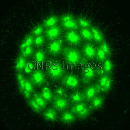
Ovocyte d'Amphioxus, "Branchiostoma lanceolatum", mesurant 140 micromètres, fixé au méthanol, observé en microscopie confocale à fluorescence. La tubuline est marquée par l'anticorps DM1A (Sigma) suivi d'un marquage secondaire couplé à la Fluorescéine. La majorité de la tubuline est maintenue sous sa forme monomérique dans des ovocytes bloqués en cycle de méiose. La perturbation de la voie de signalisation MAPK dans un ovocyte d'Amphioxus non fertilisé induit la formation d'asters cytoplasmiques résultant d'une polymérisation explosive de la tubuline. Cette image a été générée dans le cadre de contrôle de l'état monomérique de la tubuline dans des ovocytes arrêtés en méiose.
The use of media visible on the CNRS Images Platform can be granted on request. Any reproduction or representation is forbidden without prior authorization from CNRS Images (except for resources under Creative Commons license).
No modification of an image may be made without the prior consent of CNRS Images.
No use of an image for advertising purposes or distribution to a third party may be made without the prior agreement of CNRS Images.
For more information, please consult our general conditions
2015
Our work is guided by the way scientists question the world around them and we translate their research into images to help people to understand the world better and to awaken their curiosity and wonderment.