© Carlos AMPUDIA-GALVAN / Géraldine BRUNOUD / RDP / CNRS Images
Reference
20170106_0005
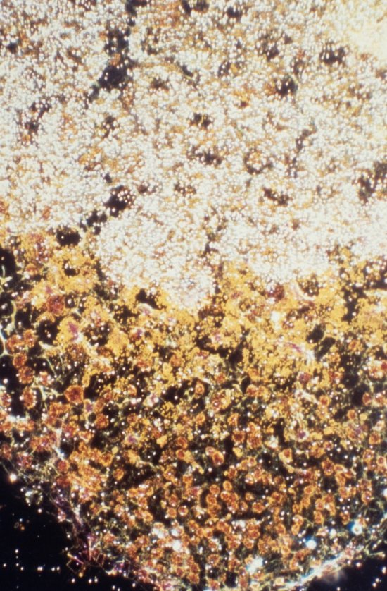
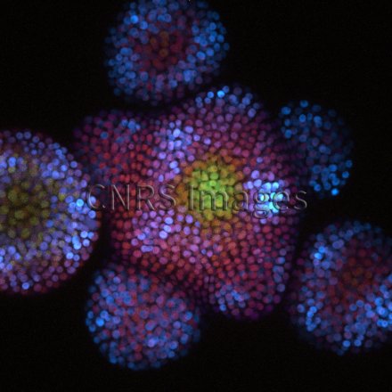
Activité hormonale dans des cellules d’Arabette des dames en microscopie confocale à fluorescence
Visual representation of hormonal activity in cells of thale cress, arabidopsis thaliana, observed using fluorescence confocal microscopy. Here, researchers are using genetically coded fluorescent markers; the level of expression and therefore hormonal activity is represented by the strength of the signal in confocal microscopy. The structure visible here is a shoot apical meristem, which produces all of a plant's new flowers (the spherical structures around the periphery).
The use of media visible on the CNRS Images Platform can be granted on request. Any reproduction or representation is forbidden without prior authorization from CNRS Images (except for resources under Creative Commons license).
No modification of an image may be made without the prior consent of CNRS Images.
No use of an image for advertising purposes or distribution to a third party may be made without the prior agreement of CNRS Images.
For more information, please consult our general conditions
