Production year
2016

© Sophie BRUSTLEIN / Rémi LASSERRE / CIML / CNRS Images
20170103_0016
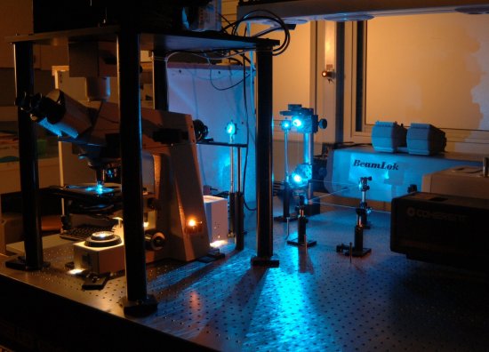
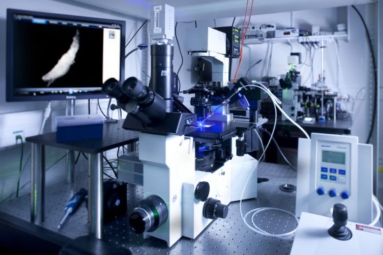
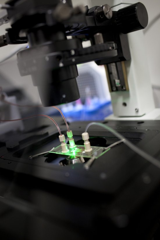
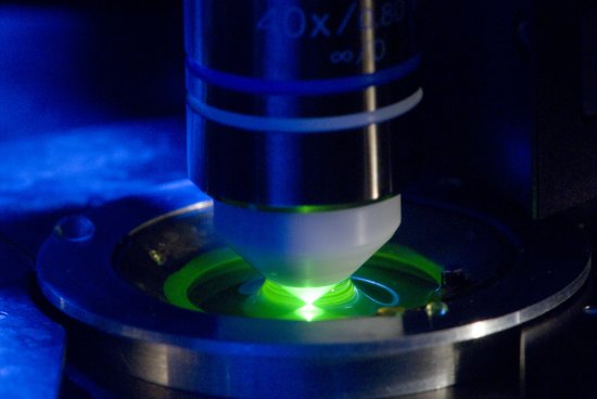
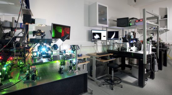
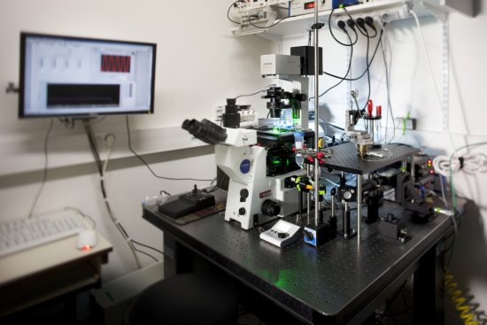
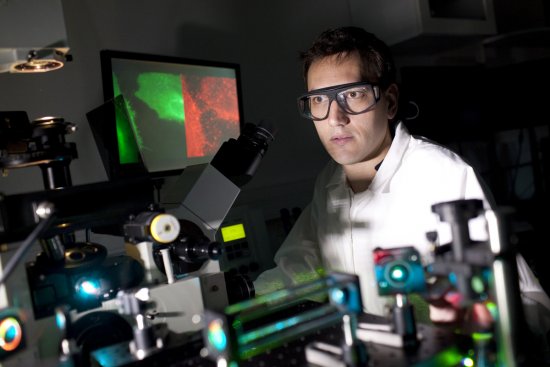
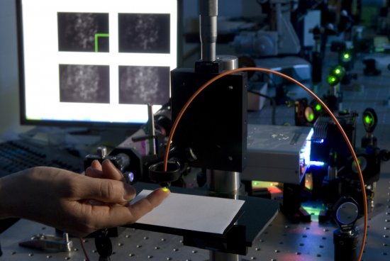
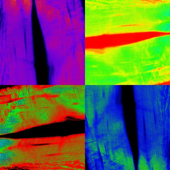
Naïve T cells (shown in green) confined in microwells (in red) filled with collagen fibres (in white). They are monitored dynamically using nonlinear microscopy. As well as the benefit of being able to image tissue in depth, nonlinear microscopy, or two-photon microscopy, reveals not only fluorescent contrasts created by marking specimens, but also additional, intrinsic contrasts that do not require marking. Unlike conventional fluorescence microscopy techniques, this technique harnesses the interaction between two or more photons and the studied material. In this case, cells are being observed by contrasting the fluorescence caused by the absorption of two photons simultaneously (CellTracker Green CMFDA). Collagen fibres are revealed by a frequency conversion process, which takes places as photons interact with an oriented, non-centrosymmetric material (second-harmonic generation). Second-harmonic generation can be used to study the three-dimensional architecture of collagen fibres, which are an essential component of the extracellular matrix, and to measure the buildup of such fibres associated with certain diseases. Lastly, microwells made of PDMS (polymer gel) have been imaged using the Coherent anti-Stokes Raman Scattering (CARS) technique, which is able to detect molecular compounds by targeting specific vibration frequencies.
The use of media visible on the CNRS Images Platform can be granted on request. Any reproduction or representation is forbidden without prior authorization from CNRS Images (except for resources under Creative Commons license).
No modification of an image may be made without the prior consent of CNRS Images.
No use of an image for advertising purposes or distribution to a third party may be made without the prior agreement of CNRS Images.
For more information, please consult our general conditions
2016
Our work is guided by the way scientists question the world around them and we translate their research into images to help people to understand the world better and to awaken their curiosity and wonderment.