© Mathieu ERHARDT / IBMP / CNRS Images
Reference
20170094_0001
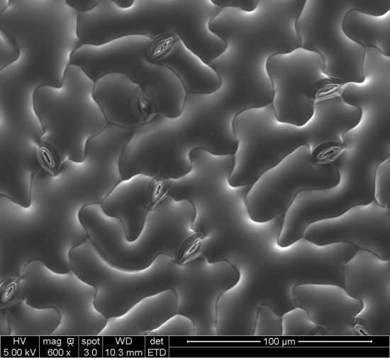
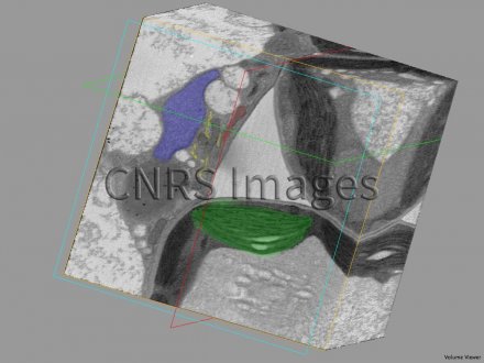
Cellules d’une feuille d’Arabette des dames reconstruite avec la technique "serial block face Imaging"
3D reconstruction of cells in a leaf of thale cress (arabidopsis thaliana), produced using the serial block face imaging technique. A total of 200 images (32 microns x 32 microns x 50 nm) were acquired using a scanning electron microscope equipped with a 3view ultramicrotome. The organelles are segmented, revealing the nucleus (in blue), the chloroplast (green), the endoplasmic reticulum (yellow) and a mitochondrion (red).
The use of media visible on the CNRS Images Platform can be granted on request. Any reproduction or representation is forbidden without prior authorization from CNRS Images (except for resources under Creative Commons license).
No modification of an image may be made without the prior consent of CNRS Images.
No use of an image for advertising purposes or distribution to a third party may be made without the prior agreement of CNRS Images.
For more information, please consult our general conditions
