© Mégane RAYER / LBCMCP / CNRS Images
Reference
20170079_0008
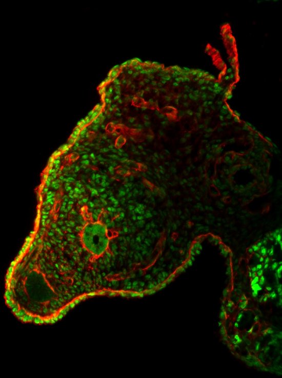
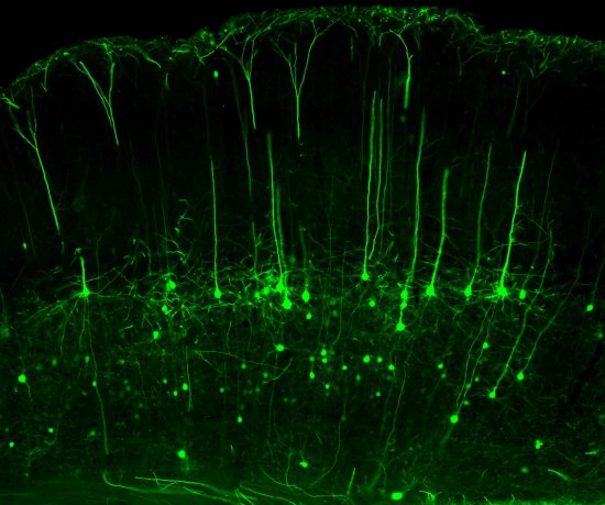
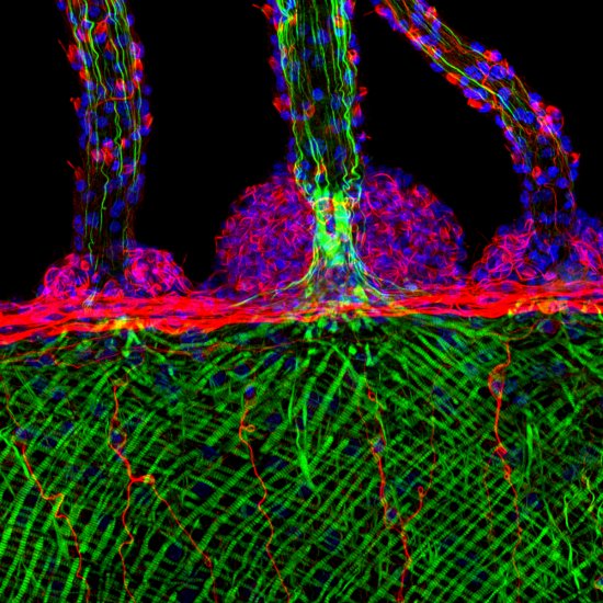
Noyaux de cellules et cytosquelette d’actine d’une patte de drosophile
Cell nuclei and actin cytoskeleton in the developing leg of a fruit fly, observed using a confocal microscope. The nuclei envelopes are visible in green, while the actin cytoskeleton is marked in red. Research scientist are testing the role of the nucleus in generating the force exercised by dying cells. This force contributes to the formation of folds, which are the origin of the future joints. More generally, the scientists aim to understand how programmed cell death or apoptosis can influence how tissues change shape.
Add to my selection
Terms of use
The use of media visible on the CNRS Images Platform can be granted on request. Any reproduction or representation is forbidden without prior authorization from CNRS Images (except for resources under Creative Commons license).
No modification of an image may be made without the prior consent of CNRS Images.
No use of an image for advertising purposes or distribution to a third party may be made without the prior agreement of CNRS Images.
For more information, please consult our general conditions