© Frank LAFONT / Sébastien JANEL / CMPI-CIIL / CNRS Images
Reference
20170071_0019
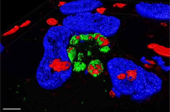
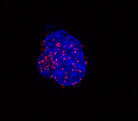
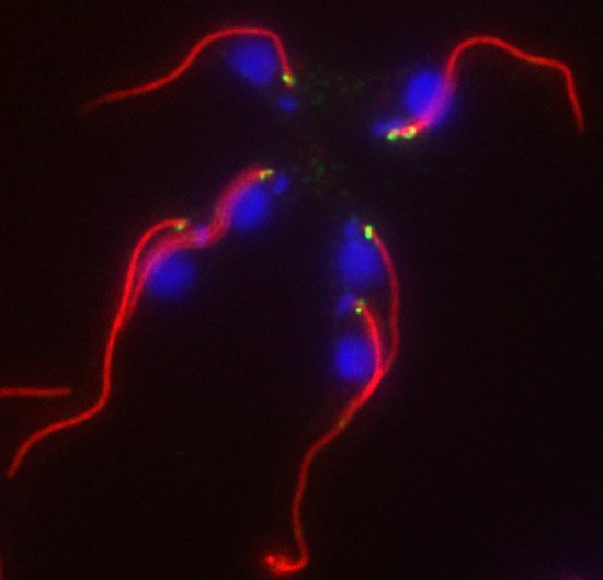
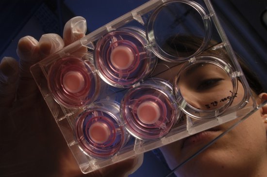
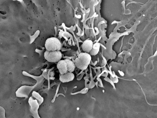
Cellules HUVEC observées en microscopie à force atomique
3D view of HUVEC endothelial cells observed using an atomic force microscope. In this view, the topology of the cell is shown on the left, and an elasticity map on the right. By manipulating this type of cell, research scientists are notably studying infection by N. meningitidis bacteria and intoxication processes involving S. aureus EDIN or anthrax EF.
Add to my selection
Terms of use
The use of media visible on the CNRS Images Platform can be granted on request. Any reproduction or representation is forbidden without prior authorization from CNRS Images (except for resources under Creative Commons license).
No modification of an image may be made without the prior consent of CNRS Images.
No use of an image for advertising purposes or distribution to a third party may be made without the prior agreement of CNRS Images.
For more information, please consult our general conditions