© Frank LAFONT / Antonio BONGIOVANNI / CMPI-CIIL / CNRS Images
Reference
20170071_0011
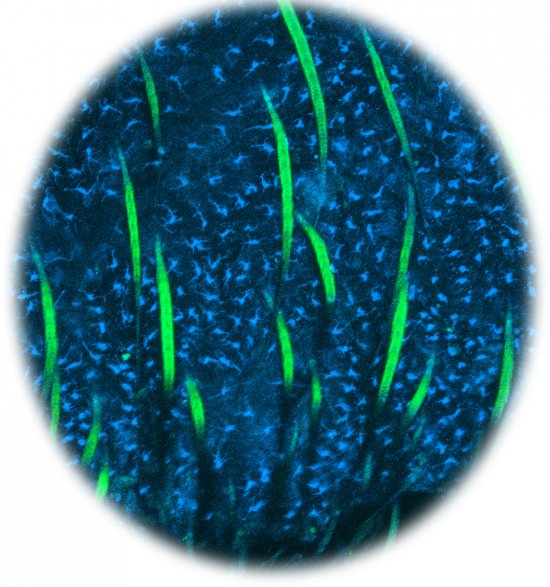
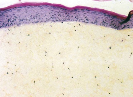
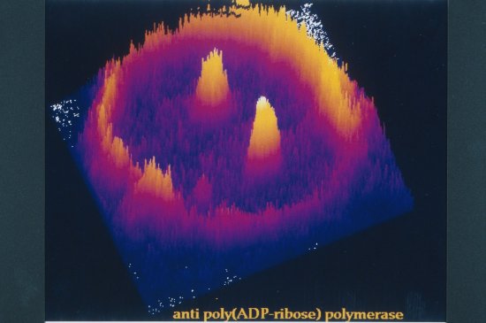
Tubuline et protéine LC3 de cellules HeLa observées en microscopie à illumination structurée
Tubulin (in green) and LC3 protein (in red) in HeLa cancer cells, observed using a structured illumination microscope. Here, the HeLa cells are being used as a model to study how the biomechanical properties of cells contribute to various processes, including infection in particular.
Add to my selection
Terms of use
The use of media visible on the CNRS Images Platform can be granted on request. Any reproduction or representation is forbidden without prior authorization from CNRS Images (except for resources under Creative Commons license).
No modification of an image may be made without the prior consent of CNRS Images.
No use of an image for advertising purposes or distribution to a third party may be made without the prior agreement of CNRS Images.
For more information, please consult our general conditions