Production year
2017

© Frank LAFONT / Nicolas BAROIS / CMPI-CIIL / CNRS Images
20170071_0007
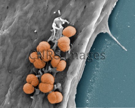
HeLa cancer cells infected with the bacterium Neisseria meningitidis, observed using a scanning microscope. The microscope image was coloured after analysis. Here, the HeLa cells are being used as a model to study how the biomechanical properties of cells contribute to various processes, including infection in particular.
The use of media visible on the CNRS Images Platform can be granted on request. Any reproduction or representation is forbidden without prior authorization from CNRS Images (except for resources under Creative Commons license).
No modification of an image may be made without the prior consent of CNRS Images.
No use of an image for advertising purposes or distribution to a third party may be made without the prior agreement of CNRS Images.
For more information, please consult our general conditions
2017
Our work is guided by the way scientists question the world around them and we translate their research into images to help people to understand the world better and to awaken their curiosity and wonderment.