Production year
2011

© Marie-Christine MIQUEL / CRCA / CNRS Images
20170055_0004
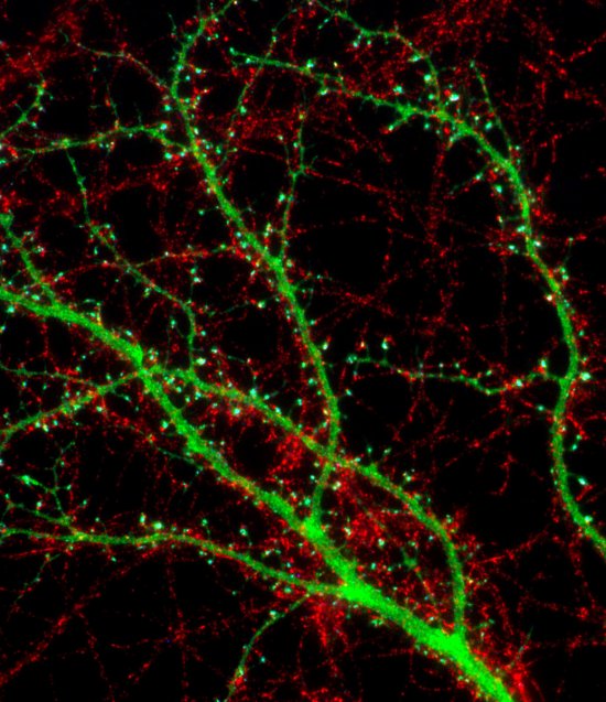
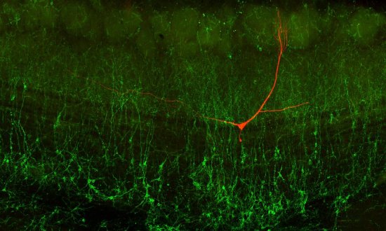
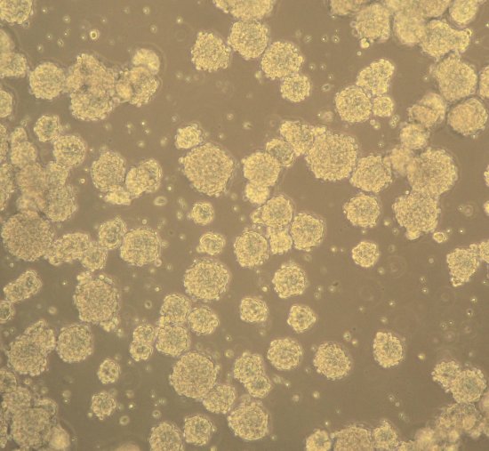
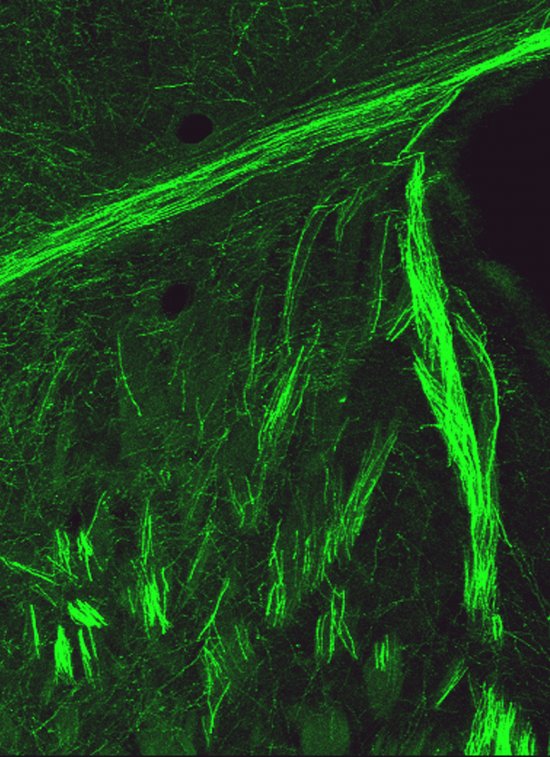
Embryonic cortical neuron from the brain of a rat cultured for nine days, viewed using a fluorescence microscope. The extensions (dendrites) of this neuron are marked in red using immunofluorescence (detection of the Map2 protein), with their mitochondria (cell electrical centres) shown in green using the fluorescent protein MitoGFP. The mitochondria form a more or less interconnected network. Their size varies depending on the neuronal compartment and the physiological and pathological context. For example, in dominant optic atrophy, a mitochondrial disease, their size is altered as well as their distribution up to the synapses. In neurodegenerative diseases such as Alzheimer’s disease, on the other hand, the morphology and functioning of the mitochondria are defective. Using different treatments adapted to the pathology, research scientists aim to mitigate neuronal malfunctions by restoring a functional mitochondrial system.
The use of media visible on the CNRS Images Platform can be granted on request. Any reproduction or representation is forbidden without prior authorization from CNRS Images (except for resources under Creative Commons license).
No modification of an image may be made without the prior consent of CNRS Images.
No use of an image for advertising purposes or distribution to a third party may be made without the prior agreement of CNRS Images.
For more information, please consult our general conditions
2011
Our work is guided by the way scientists question the world around them and we translate their research into images to help people to understand the world better and to awaken their curiosity and wonderment.