Production year
2017

© Jennifer PETERSEN/Daniel CHOQUET/IINS/CNRS Images
20170022_0004
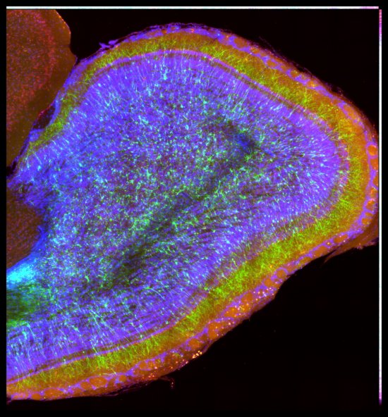
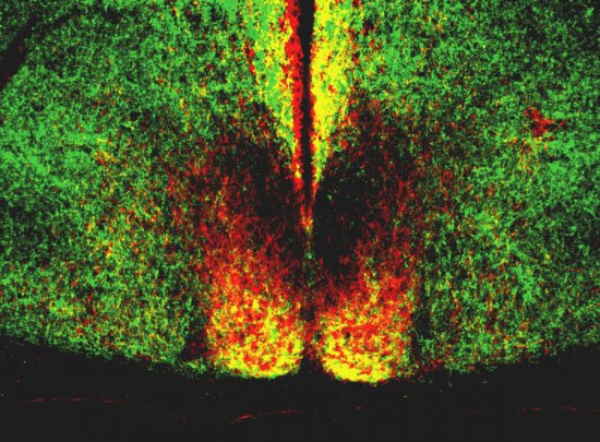
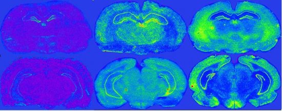
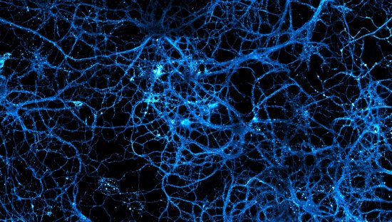
Triple marking of a rat hippocampus neuron, observed using full-field fluorescence imaging. In green, the surface marking of the GluA2 sub-unit of the AMPA-type receptors activated by glutamate. The clathrin, marked in red, enables identification of the endocytic pits that are the receptors’ transport channel to the inside of the cell. The GluA2 internalised sub-unit of the AMPA-type receptors activated by glutamate is marked in blue. A triple co-location of the clathrin, surface and intracellular receptors is revealed in way of the synapses, as is the accumulation of intracellular receptors in way of the cell body.
The use of media visible on the CNRS Images Platform can be granted on request. Any reproduction or representation is forbidden without prior authorization from CNRS Images (except for resources under Creative Commons license).
No modification of an image may be made without the prior consent of CNRS Images.
No use of an image for advertising purposes or distribution to a third party may be made without the prior agreement of CNRS Images.
For more information, please consult our general conditions
2017
Our work is guided by the way scientists question the world around them and we translate their research into images to help people to understand the world better and to awaken their curiosity and wonderment.