© Catherine SOCHA/RIDI/CNRS Images
Reference
20160113_0001
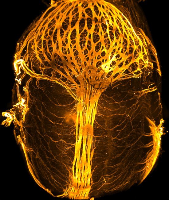
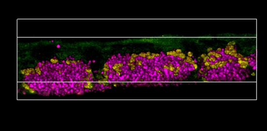
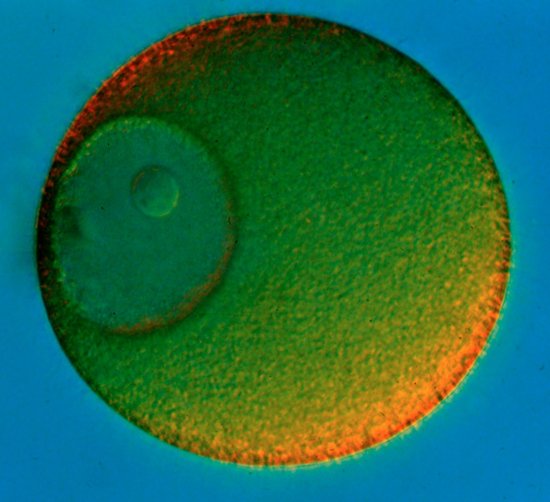
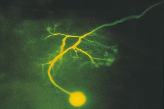
Intestinal epithelium of a drosophila infected orally by the Serratia marcescens bacterium
Intestinal epithelium of a drosophila infected orally by the Serratia marcescens bacterium. In green, a genetically marked enterocyte (intestinal cell) purges its damaged content in the lumen (inside space) of the intestine. The cell nuclei are shown in blue and the actin filaments are shown in red (at the bottom, filaments present in the visceral muscles and at the top, filament bundles below the brush border of the enterocytes). After observation of the first hours following infection by Serratia marcescens, researchers showed that the enterocytes attacked by the bacteria purge themselves rapidly of most of their cytoplasmic content. They thus rid themselves of damaged organelles, of a part of the bacteria attempting to cross the intestinal wall and apparently of the bacterial toxins. This protects them against certain harmful effects of the infection. Purging the enterocytes causes a significant but short-lived thinning of the epithelium, the layer of cells that forms the wall of the intestine. This phenomenon also occurs in humans. This work could ultimately help us improve our understanding of inflammatory diseases of the intestine such as Crohn’s disease.
Add to my selection
Terms of use
The use of media visible on the CNRS Images Platform can be granted on request. Any reproduction or representation is forbidden without prior authorization from CNRS Images (except for resources under Creative Commons license).
No modification of an image may be made without the prior consent of CNRS Images.
No use of an image for advertising purposes or distribution to a third party may be made without the prior agreement of CNRS Images.
For more information, please consult our general conditions