© Yaser HASHEM/UPR9002/CNRS Images
Reference
20160092_0001
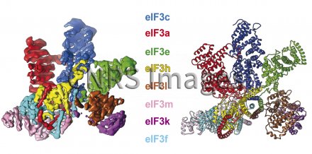
Structure of mammalian initiation factor 3 (eIF3)
Structure of mammalian initiation factor 3 (eIF3) seen by electronic cryo-microscopy (left) and interpreted by an atomic model (right). Proteins are translated from the messenger RNA by ribosome. The process is started by a crucial stage called initiation. In mammals, this stage puts more than a dozen initiation factors (eIFs) into play. Initiation factor 3 (eIF3) is comprised of 13 sub-units, 5 of which are peripheral and 8 of which are within a preserved central core, shown here. Researchers have succeeded in establishing, to a resolution of 0.6 nanometres, the three-dimensional structure of the initiation complex of mammalian protein translation. Understanding the sophisticated architecture of this complex and its interaction with key initiation factor eIF3 opens new perspectives in the search for therapeutic antiviral and anti-parasite agents.
The use of media visible on the CNRS Images Platform can be granted on request. Any reproduction or representation is forbidden without prior authorization from CNRS Images (except for resources under Creative Commons license).
No modification of an image may be made without the prior consent of CNRS Images.
No use of an image for advertising purposes or distribution to a third party may be made without the prior agreement of CNRS Images.
For more information, please consult our general conditions
