© Adrian RENGLE/Olivier BOEUF/Hélène RATINEY/Sophie CAVASSILA/CNRS Images
Reference
20110001_2014
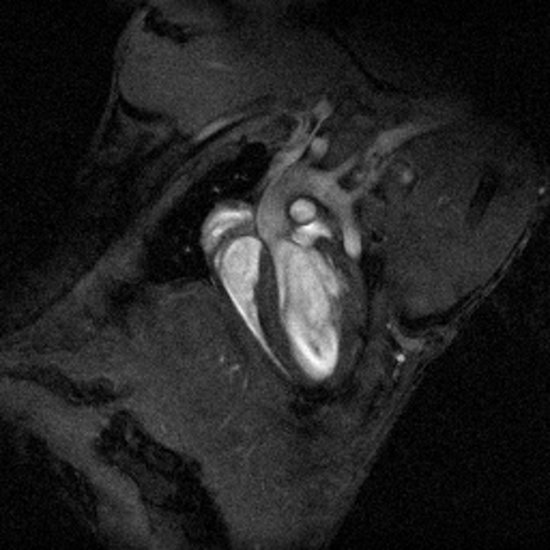
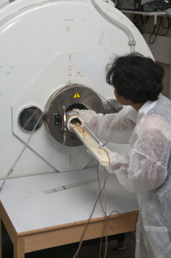
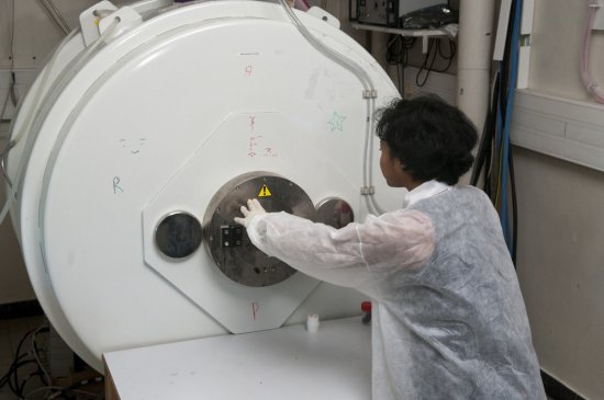
Imagerie spectroscopique du proton acquise à 4,7 tesla dans un cerveau de souris, avec un capteur mu
Imagerie spectroscopique du proton acquise à 4,7 tesla dans un cerveau de souris, avec un capteur multi-éléments à deux canaux (canal 1 : spectres rouges ; canal 2 : spectres bleus), superposée à une image anatomique en pondération T2. L'imagerie spectroscopique consiste à obtenir des images du contenu biochimique d'un organe, en réalisant l'acquisition de spectres de RMN (Résonance magnétique nucléaire) spatialement distribués.
Add to my selection
Terms of use
The use of media visible on the CNRS Images Platform can be granted on request. Any reproduction or representation is forbidden without prior authorization from CNRS Images (except for resources under Creative Commons license).
No modification of an image may be made without the prior consent of CNRS Images.
No use of an image for advertising purposes or distribution to a third party may be made without the prior agreement of CNRS Images.
For more information, please consult our general conditions