Production year
2002

© Maximilien VERMANDEL/CNRS Images
20030001_0780
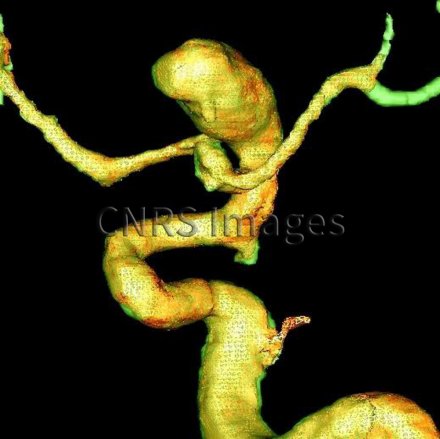
Représentation 3D d'un anévrisme de l'arbre vasculaire. Cette image résulte d'une mise en correspondance tridimensionnelle de données issues de systèmes d'imageries projectives et tomographiques d'angiographie cérébrale par rayons X (2D) et de l'imagerie par résonance magnétique (IRM). Ce recalage entre les deux modalités permet de compléter les données de l'image radiologique et de valider les innovations technologiques apportées aux dernières générations d'imageurs. Les retombées médicales de ce travail sont multiples tant dans le domaine diagnostique que thérapeutique.
The use of media visible on the CNRS Images Platform can be granted on request. Any reproduction or representation is forbidden without prior authorization from CNRS Images (except for resources under Creative Commons license).
No modification of an image may be made without the prior consent of CNRS Images.
No use of an image for advertising purposes or distribution to a third party may be made without the prior agreement of CNRS Images.
For more information, please consult our general conditions
2002
Our work is guided by the way scientists question the world around them and we translate their research into images to help people to understand the world better and to awaken their curiosity and wonderment.