Production year
1998

© CNRS Images
19990001_0450
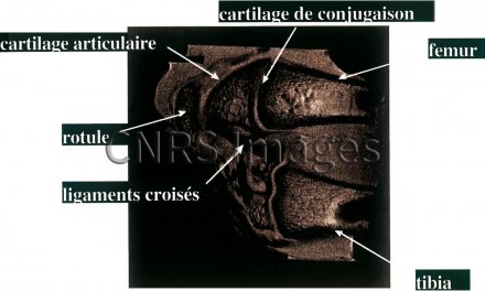
Coupe sagitale de genou de rat sain, en microscopie IRM à 8,5T ; image acquise avec une résolution de 50 microns dans le plan de l'image, et de 200 microns d'épaisseur. On reconnaît sur l'image le fémur (en haut), la rotule (en haut, à gauche), le tibia (en bas) et dans la zone articulaire, les cartilages de conjugaison et les cartilages articulaires, les deux ligaments croisés (en noir), ainsi que des détails d'organisation de l'os spongieux (dit trabéculaire).(Réf. CNRS INFO N° 367 du 15 novembre 1998).
The use of media visible on the CNRS Images Platform can be granted on request. Any reproduction or representation is forbidden without prior authorization from CNRS Images (except for resources under Creative Commons license).
No modification of an image may be made without the prior consent of CNRS Images.
No use of an image for advertising purposes or distribution to a third party may be made without the prior agreement of CNRS Images.
For more information, please consult our general conditions
1998
Our work is guided by the way scientists question the world around them and we translate their research into images to help people to understand the world better and to awaken their curiosity and wonderment.