Production year
2017

© Laurent PETIT / IMN - GIN / CNRS Images
20180066_0001
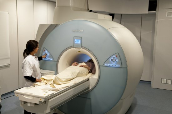
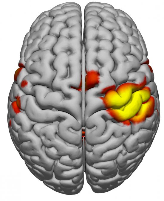
Faisceau pyramidal du cerveau humain obtenu par tractographie chez 410 sujets volontaires sains. La tractographie reconstitue les trajets des faisceaux de fibres de matière blanche du cerveau à partir des données d’IRM de diffusion. Sur cette coupe coronale passant par le sillon de Rolando, le faisceau pyramidal est constitué des axones, des cortex moteur et sensoriel reliés à la moelle épinière.
The use of media visible on the CNRS Images Platform can be granted on request. Any reproduction or representation is forbidden without prior authorization from CNRS Images (except for resources under Creative Commons license).
No modification of an image may be made without the prior consent of CNRS Images.
No use of an image for advertising purposes or distribution to a third party may be made without the prior agreement of CNRS Images.
For more information, please consult our general conditions
2017
Our work is guided by the way scientists question the world around them and we translate their research into images to help people to understand the world better and to awaken their curiosity and wonderment.