Production year
2015

© Bruno MONIER / LBCMCP / CNRS Images
20170079_0005
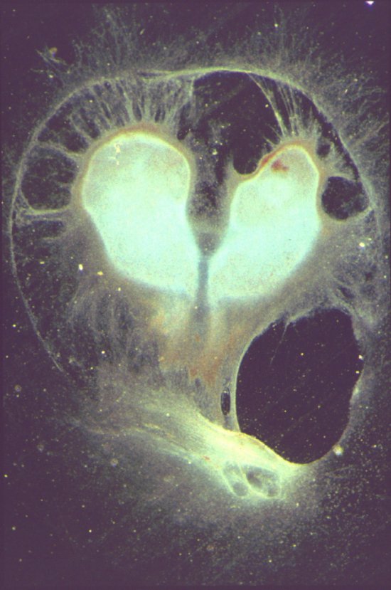
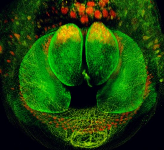
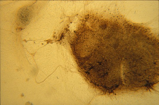
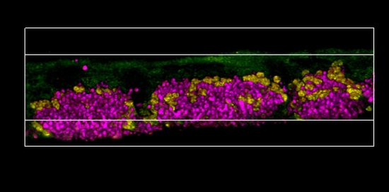
Developing fruit fly antenna observed using a confocal microscope. The aim of research scientists is to understand how programmed cell death or apoptosis can influence how tissues change shape. The image shows a developing fruit fly antenna, with the cell contours detected using an antibody directed against the E-cadherin protein (blue indicates the deepest parts, followed by green, yellow and then red, which corresponds to the surface of the sample). The presence of dying cells is visible in pink. The contours of the cells can be monitored (thanks to the display of a cell-cell adhesion molecule), making it possible to analyse the shape of the tissue and observe the presence of several concentric folds. The dying cells (located specifically at the point where future folds will form) are not eliminated passively. On the contrary, they participate actively in the formation of these folds by tugging on neighbouring cells, which results in a change in the overall shape of the tissue.
The use of media visible on the CNRS Images Platform can be granted on request. Any reproduction or representation is forbidden without prior authorization from CNRS Images (except for resources under Creative Commons license).
No modification of an image may be made without the prior consent of CNRS Images.
No use of an image for advertising purposes or distribution to a third party may be made without the prior agreement of CNRS Images.
For more information, please consult our general conditions
2015
Our work is guided by the way scientists question the world around them and we translate their research into images to help people to understand the world better and to awaken their curiosity and wonderment.