© Virginie CALLOT/CRMBM-CEMEREM/CNRS Images
Reference
20160112_0003
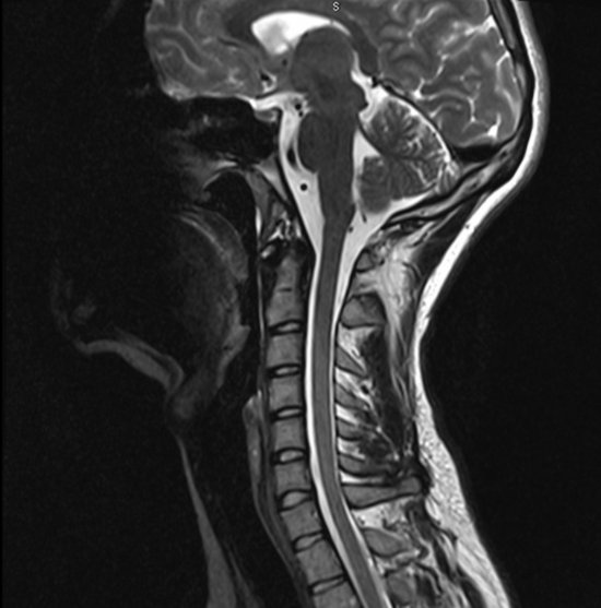
Spinal cord and spinal column, observed by multimodal conventional 3-tesla human body MRI
Spinal cord and spinal column, observed by multimodal conventional 3-tesla human body MRI, with a T1-weighted image. This imaging technique enables us to locate any anomalies in the signal which might be evidence of tissue damage. It also allows us to comprehend the geometry of the medullary canal and detect potential atrophies or degenerative changes which could, for example, induce a compression of the spinal cord.
Add to my selection
Terms of use
The use of media visible on the CNRS Images Platform can be granted on request. Any reproduction or representation is forbidden without prior authorization from CNRS Images (except for resources under Creative Commons license).
No modification of an image may be made without the prior consent of CNRS Images.
No use of an image for advertising purposes or distribution to a third party may be made without the prior agreement of CNRS Images.
For more information, please consult our general conditions