Production year
2016

© Lucie BAILLY/3SR/GIPSA-lab/LADAF/Novitom/ESRF/CNRS Images
20160111_0002
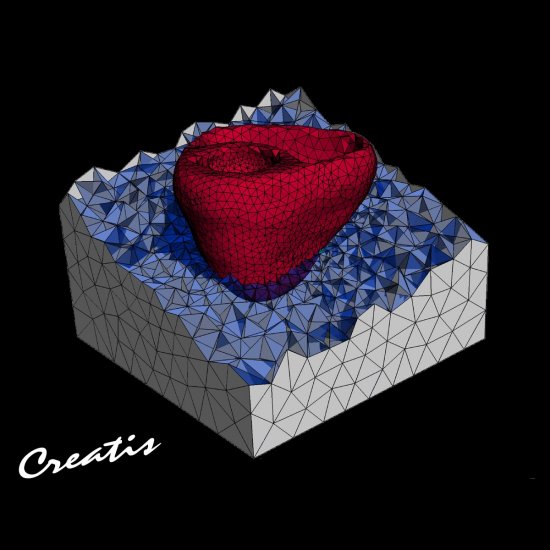
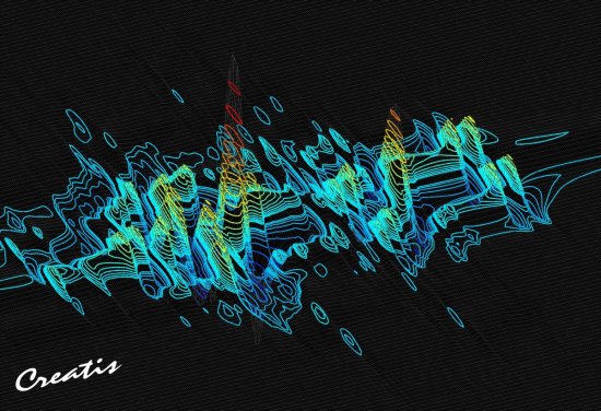
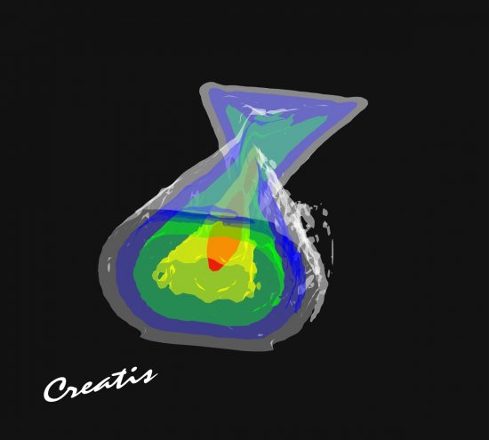
Frontal section of a human larynx, characterised by high resolution X-ray microtomography on the ESRF (European Synchrotron Radiation Facility) ID19 beamline. In the centre of the image are the two vocal folds (commonly called vocal cords); the curved structures above the vocal folds are the two vestibular folds; the uniform dark grey area at the bottom of the image is the windpipe (trachea). <br><br> Caption: Frontal section of a human larynx, characterised by high-resolution X-ray microtomography on the ESRF (European Synchrotron Radiation Facility) ID19 beamline. In the centre of the image are the vocal folds (commonly called vocal cords); the two curved structures above the vocal folds are the vestibular folds; the uniform dark grey area at the bottom is the windpipe (trachea).
The use of media visible on the CNRS Images Platform can be granted on request. Any reproduction or representation is forbidden without prior authorization from CNRS Images (except for resources under Creative Commons license).
No modification of an image may be made without the prior consent of CNRS Images.
No use of an image for advertising purposes or distribution to a third party may be made without the prior agreement of CNRS Images.
For more information, please consult our general conditions
2016
Our work is guided by the way scientists question the world around them and we translate their research into images to help people to understand the world better and to awaken their curiosity and wonderment.