Production year
2013

© Cyril FRESILLON/CNRS Images
20130001_1168
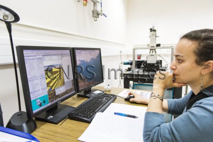
Observation d'une membrane de plusieurs feuillets de graphène, grâce à un dispositif de spectroscopie Raman couplée à des mesures de conductance. Un logiciel permet de positionner le spot laser servant de sonde. La membrane est attachée à une extrémité par une électrode d'or déposée sur silice, qui fait vibrer la membrane par application d'un potentiel électrostatique. Le dispositif permet des mesures en réflectométrie, en Raman et en fluorescence, si nécessaire. Il s'agit d'un microscope confocal très lumineux utilisé pour la cartographie Raman spatialement résolue. Cela permet notamment de mesurer les vibrations de la membrane grâce à la lumière. De faibles quantités de molécules peuvent être détectées grâce à ces membranes.
The use of media visible on the CNRS Images Platform can be granted on request. Any reproduction or representation is forbidden without prior authorization from CNRS Images (except for resources under Creative Commons license).
No modification of an image may be made without the prior consent of CNRS Images.
No use of an image for advertising purposes or distribution to a third party may be made without the prior agreement of CNRS Images.
For more information, please consult our general conditions
2013
Our work is guided by the way scientists question the world around them and we translate their research into images to help people to understand the world better and to awaken their curiosity and wonderment.