© Olivier COLLIOT/CNRS Images
Reference
20080001_0490
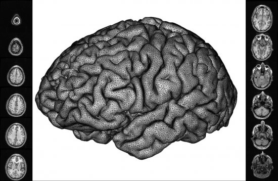
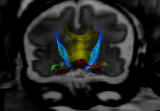
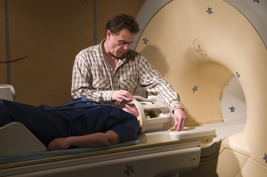
Imagerie cérébrale : à gauche, hippocampe d'un sujet sain, à droite hippocampe d'un patient atteint
Imagerie cérébrale : à gauche, hippocampe d'un sujet sain, à droite hippocampe d'un patient atteint de la maladie d'Alzheimer. Grâce au logiciel de traitement d'images mis au point par des chercheurs, un radiologue peut avoir accès au volume de l'hippocampe, représenté en rouge sur la coupe. L'hippocampe est une structure du cerveau jouant un rôle fondamental dans les processus de mémoire ; il est affecté aux premiers stades de la maladie d'Alzheimer. Ce logiciel a été utilisé avec succès pour distinguer des patients atteints par la maladie d'individus en bonne santé et de même âge.
Add to my selection
Terms of use
The use of media visible on the CNRS Images Platform can be granted on request. Any reproduction or representation is forbidden without prior authorization from CNRS Images (except for resources under Creative Commons license).
No modification of an image may be made without the prior consent of CNRS Images.
No use of an image for advertising purposes or distribution to a third party may be made without the prior agreement of CNRS Images.
For more information, please consult our general conditions