© Jean-René MARTIN/CNRS Images
Reference
20070001_0522
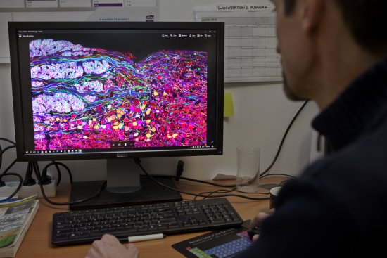
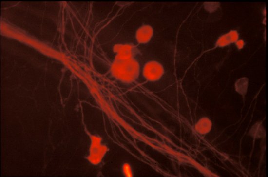
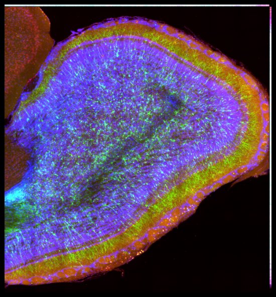
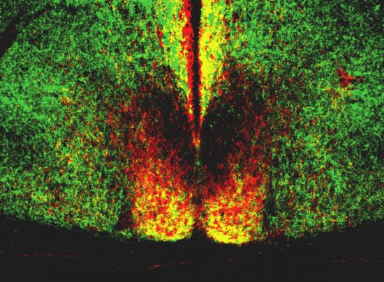
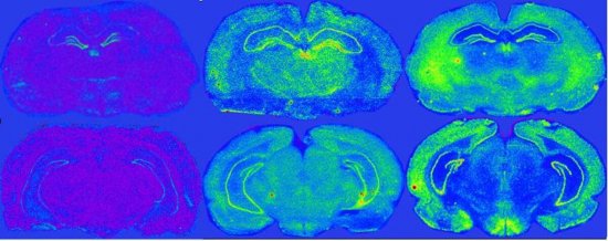
Nouvelle technique d'imagerie neuronale fonctionnelle chez la drosophile. A gauche : image micro-pho
Nouvelle technique d'imagerie neuronale fonctionnelle chez la drosophile. A gauche : image micro-photographique prise en fluorescence (à partir de la GFP), des corps pédonculés, avec en superposition, un dessin schématique des différentes parties des corps pédonculés. A l'arrière et en haut, en rouge : les corps cellulaires (cell bodies), en jaune, le calyx (correspondant aux dendrites). A l'avant, les lobes (correspondant aux projections ou axones) en rouge (lobes a / ß), en vert (lobes a'/ß') et en bleu (lobes y). Bar-échelle : 50 µM. A droite : image en bioluminescence, représentant la réponse secondaire retardée (d'environ 10 minutes), suite à la stimulation par la nicotine (un agoniste de l'acétylcholine, mimant ainsi un input olfactif).
Add to my selection
Terms of use
The use of media visible on the CNRS Images Platform can be granted on request. Any reproduction or representation is forbidden without prior authorization from CNRS Images (except for resources under Creative Commons license).
No modification of an image may be made without the prior consent of CNRS Images.
No use of an image for advertising purposes or distribution to a third party may be made without the prior agreement of CNRS Images.
For more information, please consult our general conditions