© Chantal GUIDI RONTANI/CNRS Images
Reference
20010001_0292
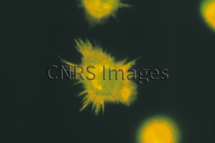
Micrographie d'un macrophage péritonéal de souris infecté par "Bacillus anthracis" (agent de la mala
Micrographie d'un macrophage péritonéal de souris infecté par "Bacillus anthracis" (agent de la maladie du charbon ou "anthrax"). Les spores phagocytées puis germées sont révélées par fluorescence à l'aide d'un marquage indirect par des anticorps de lapin anti-bacille et des anticorps de chèvre anti-IgG de lapin marqués (en jaune). Les macrophages sont visualisés après marquage de leurs filaments d'actine par la phalloidine marquée (en vert).
The use of media visible on the CNRS Images Platform can be granted on request. Any reproduction or representation is forbidden without prior authorization from CNRS Images (except for resources under Creative Commons license).
No modification of an image may be made without the prior consent of CNRS Images.
No use of an image for advertising purposes or distribution to a third party may be made without the prior agreement of CNRS Images.
For more information, please consult our general conditions
