© Rémi DUMOLLARD / LBDV / MICA / CNRS Images
Reference
20180020_0003
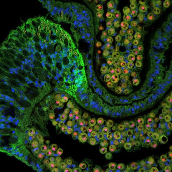
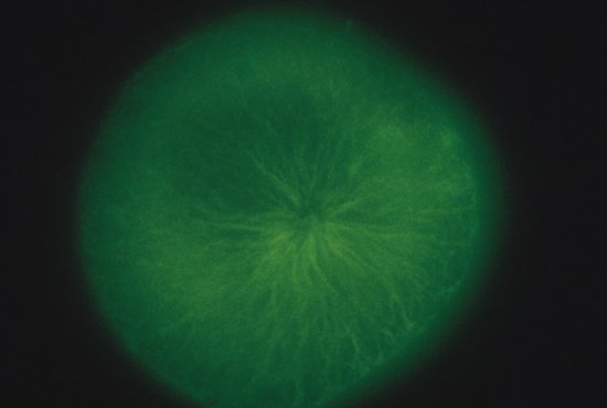
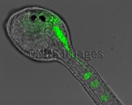
Embryon d'Ascidie blanche observé en microscopie à fluorescence
Embryon d'Ascidie blanche, "Phallusia mammillata", observée en microscopie à fluorescence plein champ, injecté au stade de 16 cellules dans le blastomère du lignage neural et notochorde avec un mRNA (ARN Messager) codant pour MAP7 :: GFP . En vert, on observe le MAP7 (utilisé pour visualiser les microtubules dans des cellules vivantes).
The use of media visible on the CNRS Images Platform can be granted on request. Any reproduction or representation is forbidden without prior authorization from CNRS Images (except for resources under Creative Commons license).
No modification of an image may be made without the prior consent of CNRS Images.
No use of an image for advertising purposes or distribution to a third party may be made without the prior agreement of CNRS Images.
For more information, please consult our general conditions

