Production year
2014

© Maeva ORLIAC/ISEM/CNRS Images
20140001_1459
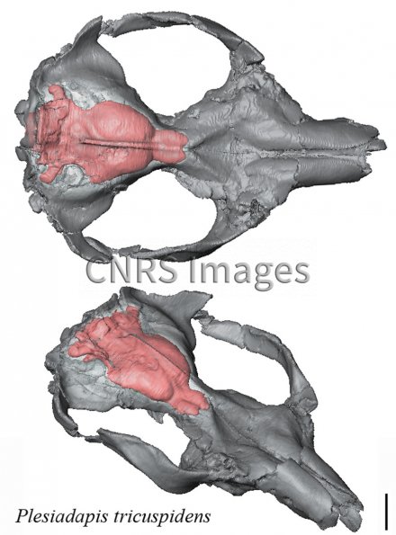
Reconstitution 3D in situ du moulage endocrânien de "Plesiadapis tricuspidiens". Celle-ci a été obtenue grâce à un microtomographe à rayons X, à partir d'un crâne fossilisé de "Plesiadapis" provenant des collections du Muséum d'histoire naturelle. "Plesiadapis" est un petit primate ayant vécu entre 58 et 52 millions d'années. C'est un proche cousin des "primates vrais", le groupe auquel l'homme et les grands singes appartiennent. Les chercheurs sont parvenus à reconstituer la forme et l'apparence du cerveau grâce à des marques laissées sur la surface interne du crâne. Ils ont constaté que "Plesiadapis" possédait un cerveau peu volumineux. Mais l'étude de sa structure a montré qu'elle préfigurait celle du cerveau des "primates vrais". Les modifications structurelles de l'encéphale des primates étaient donc apparues avant même que leur taille n'augmente.
The use of media visible on the CNRS Images Platform can be granted on request. Any reproduction or representation is forbidden without prior authorization from CNRS Images (except for resources under Creative Commons license).
No modification of an image may be made without the prior consent of CNRS Images.
No use of an image for advertising purposes or distribution to a third party may be made without the prior agreement of CNRS Images.
For more information, please consult our general conditions
2014
Our work is guided by the way scientists question the world around them and we translate their research into images to help people to understand the world better and to awaken their curiosity and wonderment.