Production year
2017

© Christophe HARGOUES / Institut de la Vision / CNRS Images
20170008_0055
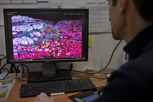
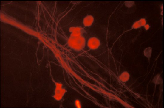
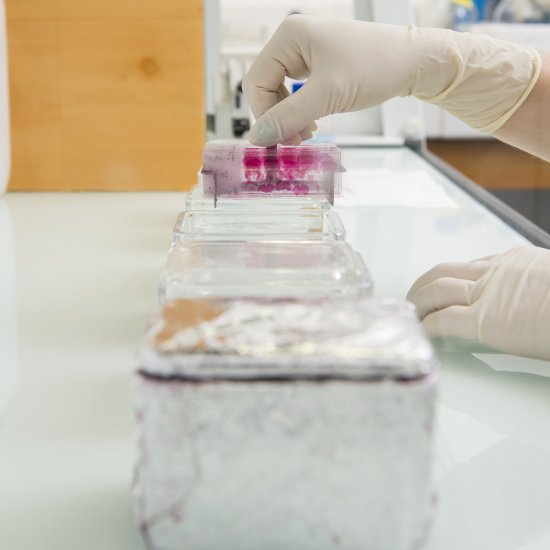
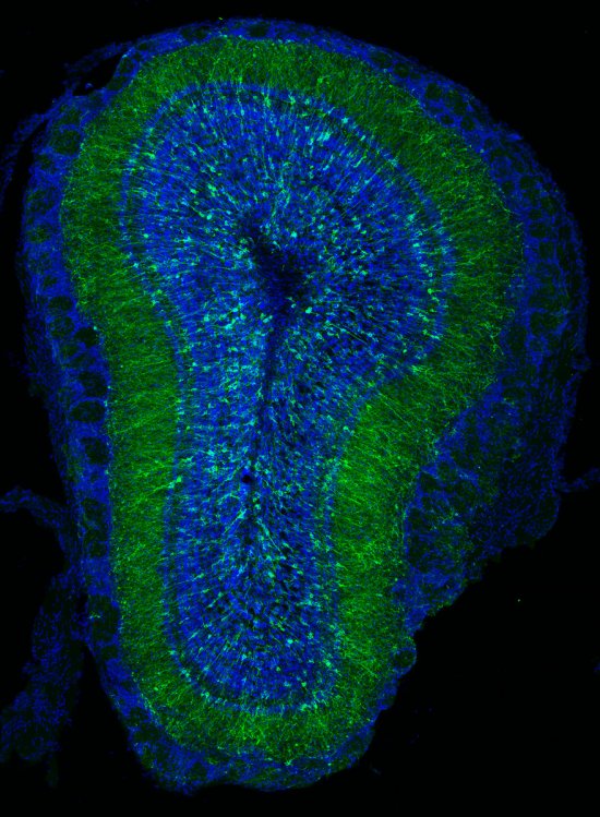
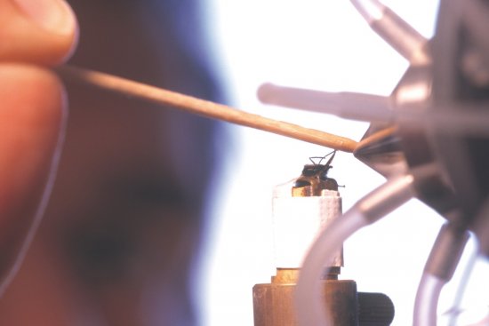
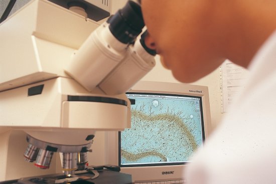
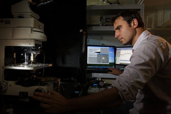
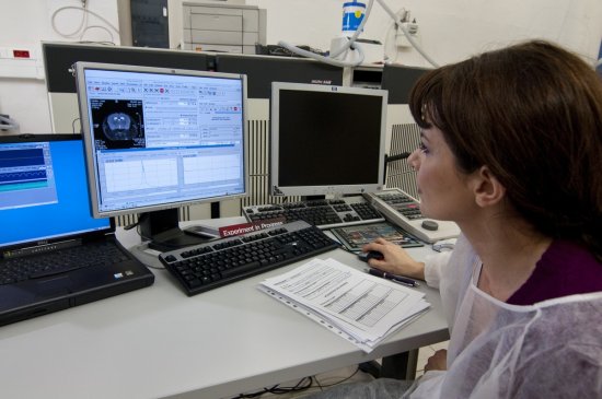
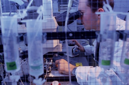
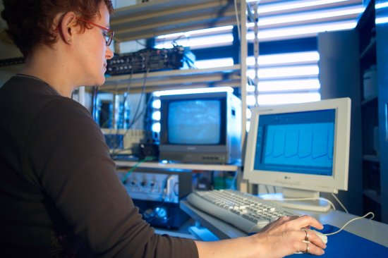
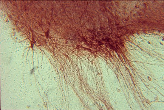
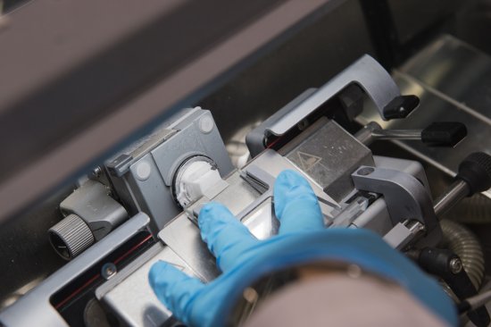
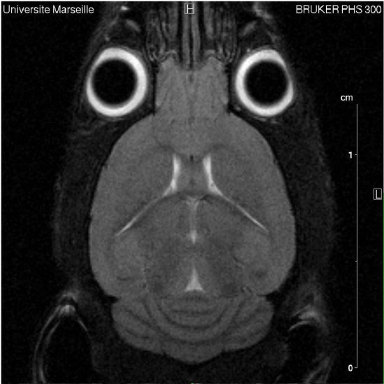
Mouse embryo placed in a carrier to then observe it under a selective plane illumination microscope. This technique enables rapid 3D imaging of specimens (here the organisation of the brain and the visual pathways). The goal is to see how the axons are positioned in this embryo.
The use of media visible on the CNRS Images Platform can be granted on request. Any reproduction or representation is forbidden without prior authorization from CNRS Images (except for resources under Creative Commons license).
No modification of an image may be made without the prior consent of CNRS Images.
No use of an image for advertising purposes or distribution to a third party may be made without the prior agreement of CNRS Images.
For more information, please consult our general conditions
2017
Our work is guided by the way scientists question the world around them and we translate their research into images to help people to understand the world better and to awaken their curiosity and wonderment.